


RECENT PUBLICATIONS: Improvement of Bovine Nucleus Pulposus Cells Isolation Leads to Identification of Three Phenotypically Distinct Cell Subpopulations
INEB researchers recently published an article in the journal Tissue Engineering Part A, available since August 2015. The article is entitled "Improvement of Bovine Nucleus Pulposus Cells Isolation Leads to Identification of Three Phenotypically Distinct Cell Subpopulations" and is authored by Maria Molinos, Catarina R. Almeida, Raquel M. Gonçalves, and Mário A. Barbosa.
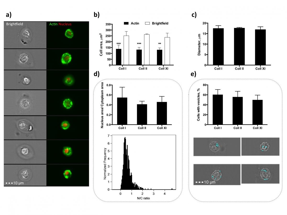
Fig.2 Morphological analysis of bNP cells. (a) Images of bNP cells obtained by Imaging Flow Cytometry (first column: brightfield images of cells; second column: merged fluorescence images of cells stained with phalloidin-FITC (actin, green) and DRAQ5 (nuclei, red); (b) bNP cell area based on brightfield (white bars) and actin staining (black bars) images of tissue digested with different collagenases: Coll-I, Coll-II, and Coll-XI (2.0 mg/mL, 4 h digestion); (c) bNP cell diameter based on actin staining for tissue digested with the three collagenases; (d) nuclear-to-cytoplasmic (N/C) ratio of bNP cells isolated with the three different methods (top), and representative histogram of N/C ratio distribution of bNP cells isolated with Coll- I, 2.0 mg/mL for 4 h (bottom); (e) Percentage of cells containing one or more vesicles after isolation with the different collagenases—a light blue mask specifically designed to mark and quantify bright vesicles within cells is shown in the bottom brightfield images of cells. Results are presented as mean – SD of three independent experiments, where a total of 3661 cells for Coll-I, 993 cells for Coll-II, and 5385 cells for Coll-XI were analyzed. In the case of cell area analysis, groups were compared using a two-way ANOVA (Bonferroni’s multiple-comparisons test). **0.001 < p < 0.01 and ***p < 0.001. Color images available online at www.liebertpub.com/tea
This study establishes an improved method for bovine NP (bNP) cell isolation, whose procedure is still not consensual among the literature, allowing a thorough characterization of cell (sub)populations that exist in the young NP. It was found that bNP cells present a similar morphology independently of the digestive enzyme used. However, cell yield was greatly improved by Coll-XI (2 mg/mL) treatment for a short digestion period. Interestingly, three subpopulations, with different sizes and auto-fluorescence, were consistently identified by flow cytometry. And crucially, differential expression of cell markers was found among these subpopulations. Such knowledge is key for a better understanding of NP cell biology and its potential endogenous regenerative capacity.